Texto principal:
La isquemia macular diabética se caracteriza por la disminución de la vasculatura foveal, lo que causa daño en la red capilar. Dicha patología es una causa importante de discapacidad visual. La patología provoca pérdida de tejido neuroretiniano y daño funcional precoz debido a hipoxia o hiperglucemia. La isquemia persistente de la red de capilares maculares superficiales y profundos puede ocasionar daño neurosensorial permanente y por lo tanto provocar nuevos cambios microvasculares.
La angiografía fluoresceínica es un procedimiento utilizado para el diagnóstico de la isquemia macular diabética. Dicha técnica muestra grandes zonas de hipofluorescencia debido a la ausencia capilares. El Estudio para el tratamiento de la retinopatía diabética (ETDRS) recomienda la angiografía fluoresceínica como técnica para evaluar la gravedad del daño isquémico.
La angiografía mediante tomografía de coherencia óptica es un nuevo método de estudio basado en imágenes de alta resolución que muestran la circulación retiniana y coroidal sin sustancia de contraste. Este método confiable, en tiempo real y no invasivo permite visualizar las zonas no irrigadas en la región macular asociadas a la retinopatía diabética.
La capa interna de la macula está formada por una capa de fibras nerviosas retinianas, una capa de células ganglionares y una capa plexiforme interna que forma el complejo celular ganglionar. La integridad de los capilares es fundamental para la supervivencia de las células ganglionares. El estado del complejo de células ganglionares es indicador de la gravedad de la isquemia macular diabética.
El objetivo del presente estudio fue evaluar la eficacia de la angiografía mediante OCT y compararla con la angiografía fluoresceínica para el diagnóstico de la retinopatía diabética isquémica y su clasificación según la zona avascular foveal (ZAF).
Pacientes y métodos: Participaron 20 pacientes (31 ojos) con maculopatía diabética isquémica 17 controles. Tanto los pacientes como los controles fueron sometidos a examen oftalmológico completo, incluyendo agudeza visual, presión intraocular, angiografía fluoresceínica y angiografía mediante TCO. Las imágenes de la angiografía fluoresceínica y de la angiografía mediante TCO fueron clasificadas según la zona avascular. El espesor del complejo de células ganglionares se evaluó mediante TCO.
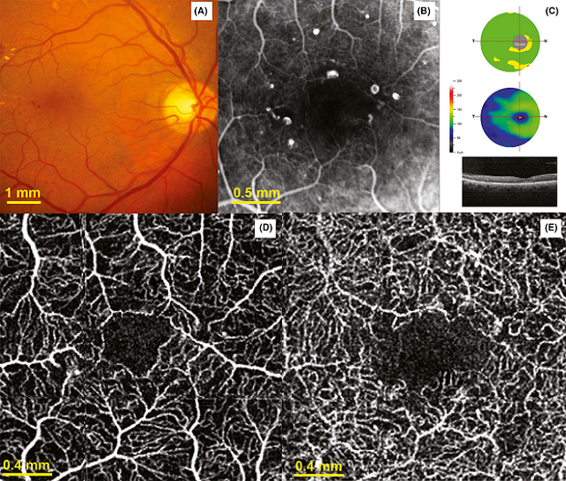
ETDRS. Maculopatía diabética isquémica grado 2. A) fotografía de fondo de ojo en color. B) Angiografía fluoresceínica que muestra ampliación de la zona avascular foveal. C) Reducción del complejo de células ganglionares en el mapa de significancia y en el espesor. D, E) Angiografía mediante TCO del plexo capilar superficial y profundo que muestra capilares truncados y red vascular perifoveal irregular.
Para evaluar si la angiografía por TCO podría ser una alternativa no invasiva a la angiografía fluoresceínica, utilizamos los estándares del ETDRS para clasificar las imágenes obtenidas y encontramos una buena correlación entre ambos procedimientos. Asimismo, las zonas de isquemia estudiadas mediante angiografía por TCO fueron mucho más definidas que con la angiografía fluoresceínica dado que no hay velamiento causado por filtración de tintura. Especialmente, la angiografía fluoresceínica no llega a mostrar imágenes de los capilares profundos.
Por otro lado, en la angiografía por TCO, se pueden identificar alteraciones en el tamaño de los vasos, flujo y morfología al examinar las imágenes de los plexos capilares superficial y profundo. Las imágenes de TCO muestran claramente pocos vasos residuales en el plexo superficial con capilares truncados y en el plexo profundo, ramificaciones anastomoticas verticales que unen el plexo superficial y profundo en la maculopatía diabética isquémica.
Además, se han identificado las alteraciones histopatológicas de la retina interna que provocan la pérdida de células ganglionares en pacientes con retinopatía diabética. Confirmando lo observado en estudios anteriores, en el presente estudio el espesor del complejo de células ganglionares se vio reducido significativamente, pero sin ninguna correlación con la agudeza visual.
Esto se contrapone a lo publicado en nuestro estudio anterior donde se observó una correlación entre agudeza visual, espesor del complejo de células ganglionares y pérdida de sensibilidad retiniana en ojos con dicha patología. (Cennamo et al. 2015). Esta discrepancia puede surgir de la mayor gravedad de la patología en los pacientes del estudio anterior. Los métodos anteriores se basan en complejos modelos matemáticos para cuantificar el daño isquémico, en cambio la TCO lo calcula automáticamente tanto en imágenes superficiales como profundas.
La limitación de la angiografía por TCO es que el paciente debe fijar con precisión durante varios segundos, mientras que la angiografía fluoresceínica se obtiene en fracción de segundo, junto con información sobre flujo sanguíneo e información fisiológica sobre la salud de los vasos.
Conclusiones:
Los resultados del presente estudio indican que la angiografía por TCO es un método válido, confiable y sencillo para evaluar y clasificar el daño de la red capilar sin administrar medio de contraste.
Resumen y comentario objetivo: Dr. Martín Mocorrea